Frequently Asked Questions
Can I have a CT scan and a nuclear scan done in one day ?
The next question friends often ask is whether we have undergone a CT scan or MRI, or if we plan to do a CT scan and MRI. Do these two procedures interfere with nuclear medicine or not?
We divide this issue into two parts: first, the patient visits nuclear medicine and has their scan done. After that, there is no problem from our side for them to undergo a CT scan or MRI. However, since a radiopharmaceutical agent has been injected into the patient, it is better to delay this procedure for 24 hours so that the radioactive material does not come into contact with pregnant women or individuals under 4 to 5 years old, and radiation does not affect these individuals. But from a technical standpoint, there is no problem in this regard.
The next point is that if friends have undergone a CT scan, MRI, or nuclear scan 24 to 48 hours before the nuclear medicine procedure, they should definitely inform us. Because if a contrast-enhanced CT scan with oral contrast has been performed, at least 48 hours should have passed since the CT scan. Only then can we proceed with the nuclear imaging.
For those who have had an MRI, there are no restrictions or limitations. Therefore, they can easily proceed with the nuclear medicine procedures afterward. Thank you for your cooperation.
What is a CT scan?
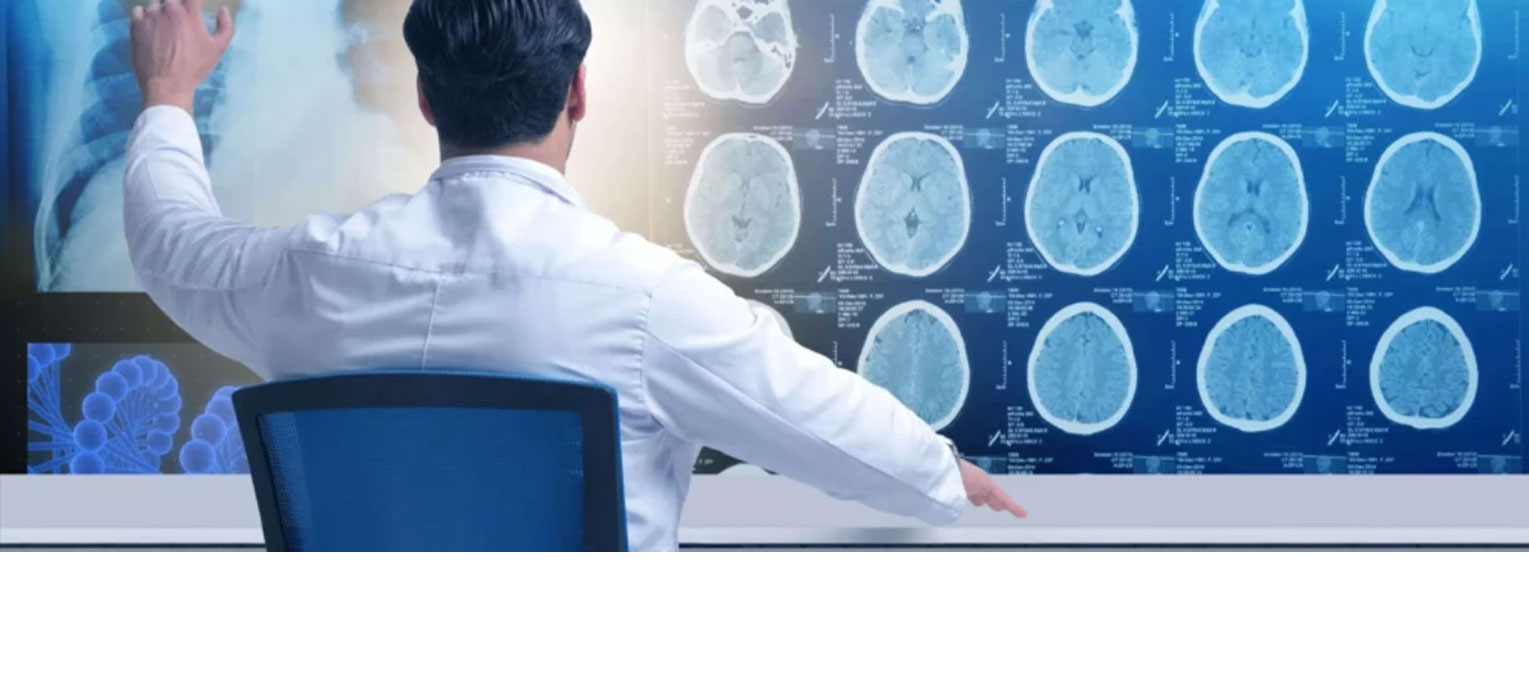
Computed tomography or CT scan is an advanced medical imaging technique often prescribed by doctors. X-rays and CT scans have greatly contributed to the medical community. Today, CT scans are an indispensable part of diagnosis and treatment. The internal tissues of the body can be visualized with this imaging method. As a result, specialists can examine their shape and potentially identify possible diseases. The image produced by a CT scan shows cross-sectional views of the body's organs.
What is MRI?
Most specialists prescribe MRI for better visualization of the body's condition. This test has also been a significant service to the medical community. Many patients feel a bit anxious before undergoing this test because they have to lie on a bed and enter a tunnel for imaging. But there is no need to worry at all. No discomfort or stress will be caused inside the tunnel.
MRI stands for Magnetic Resonance Imaging. This method uses a magnetic field for imaging, providing precise images of body parts. This magnetic field can align water molecules in the body. Radio waves then interact with these aligned atoms, producing weak signals. The MRI images are generated using this energy in the system.
I have a prosthesis in my body. Can I undergo nuclear imaging?
Certainly! Here's a more professional and polished translation of your text:
Some visitors have raised two common questions. The first concerns whether the presence of prosthetic devices—such as pacemakers, hearing aids, knee prostheses, dental implants, etc.—within the body can influence the results of nuclear imaging scans. The second pertains to whether these devices could be affected by the imaging process, potentially impairing their functionality.
Rest assured, dear visitors
Since nuclear medicine imaging devices do not generate magnetic fields, this type of imaging is entirely safe for individuals with prostheses. Patients can undergo the procedure confidently without concern. We appreciate your trust and confidence in our services.
What is a Prosthesis?
In medical terminology, prostheses refer to artificial limbs, members, or orthopedic devices. Broadly speaking, a prosthesis is an external object placed within or on the body for cosmetic or therapeutic purposes. Many individuals have devices such as pacemakers, knee prostheses, dental prostheses, and more. Naturally, they may be concerned about whether these devices could be affected by nuclear imaging procedures.
What is the difference between nuclear scan and CT angiography of the heart?
Myocardial nuclear scanning shows the blood supply to the heart muscle. It indicates whether the artery behind this muscle, responsible for supplying blood to it, is blocked or what its condition is.
CT angiography reveals the anatomy of the coronary blood vessels. It shows how much narrowing or blockage exists in these vessels (not in the heart tissue itself).
The state of the nuclear scan is that** the artery behind the muscle may be blocked, and the muscle receives blood from the neighboring arteries. Even if the artery responsible for blood supply is obstructed, the muscle continues to function by receiving blood from adjacent vessels, and the heart tissue remains well-perfused and capable of continuing its activity—unless during intense exertion, when the muscle may become ischemic. In such cases, the patient is referred for revascularization procedures.
What is CT Angiography?
It is a modern method used to diagnose the anatomy of the coronary blood vessels. This technique involves the use of a CT scanner and a special contrast agent to enhance the images of the vessels.
This contrast agent is injected intravenously, typically into the arm, and allows imaging of all body parts. Using X-ray and computer technology, cross-sectional images of the inside of the body are produced.
If the specialist detects abnormalities in various vessels, they may prescribe a CT angiography for the patient. The information obtained from this imaging helps the doctor better diagnose the patient's condition.
Applications of CT Angiography include:
- Detecting vessel inflammation (aneurysm)
- Blockages
- Blood clots
- Congenital cardiovascular anomalies
- Vascular defects such as system malformations
- Various injuries
- Benign and malignant tumors
- Vessel rupture or tearing